| welcome | research | publications | vita |
Research
Publications -- fMRI Research -- Electrophysiology
|
Keywords: fMRI - Visual Cortex - Retinotopic Mapping - Cognitive Neurology
|
|
fMRI Research
|
|
|
|
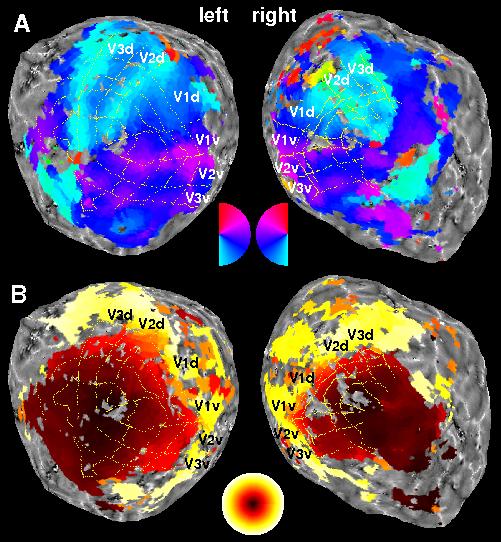
Retinotopic Mapping Retinotopic maps overlayed on flattened representations of the occipital poles of a healthy volunteer. (A) Colors indicate the underlying functional voxels' preference for the polar angle of visual stimuli. The systematic color changes across the visual cortex reflect the retinotopic organization of early visual areas (V1-V3). (B) Functional voxels' preference for the eccentricity of visual stimuli. |
|
|
|
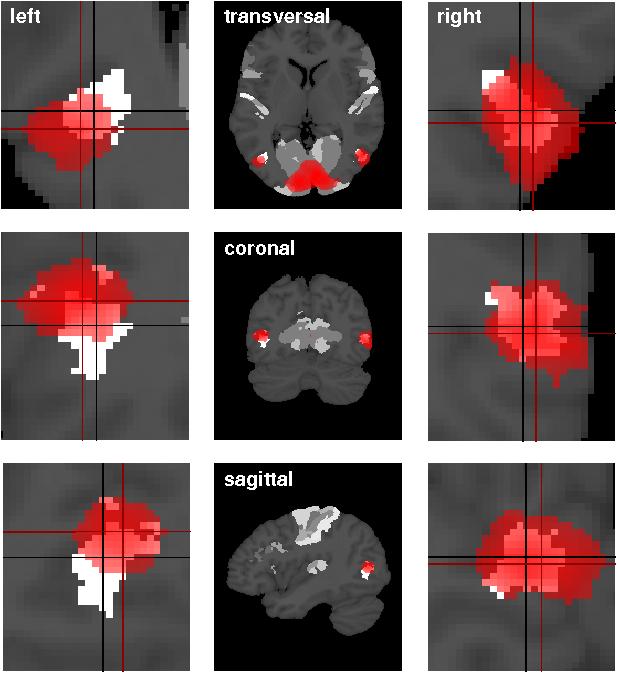
Functional validation of cytoarchitectonic maps The cytoarchitectonic delineation of cortical areas in human postmortem brains allows the construction of probability maps for anatomically defined cortical areas. In order to use these maps for the assignment of cortical activations in functional imaging studies, each anatomical map has to be functionally validated. In the above example, using fMRI, we have identified putative human area V5/MT+ in normals by modelling the BOLD responses to alternating radially moving and stationary dot patterns. Functional activations were compared to cytoarchitectonic probability maps (aPM) of its putative correlate area hOc5 which was calculated based upon data from histological sections of 10 human post-mortem brains. Bilateral visual cortex activations were seen in the single subject dynamic vs. stationary contrasts and in the group random-effects analysis. Comparison of area hOc5 with functional probability maps (in red) showed that most of area hOc5 was covered by the respective fPM. The correspondence between the functionally defined human V5/MT+ and the post-mortem cytoarchitectonic area hOc5 suggests that the anatomically defined area hOc5 can be used for a more informed analysis of functional imaging data. |